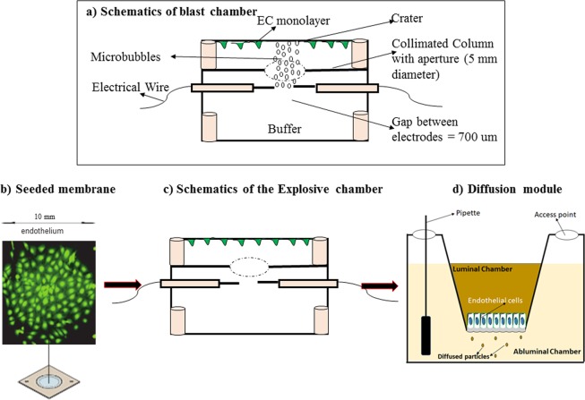
Figure 1.
Schematics of the custom-designed blast chamber and a brief flow of experimental protocol from culture insert, proof of cell adhesion to PETE membrane to the blast chamber and finally the diffusion chamber. (a) The blast chamber was engineered to generate shockwave-induced microbubbles. They can only rise to the top of the chamber and collapse onto the seeded BECs, detaching cells from a controlled area referred to as a “crater”. (b) Cell culture insert. Green FITC cell tracker was used to demonstrate that the PETE membrane coated with fibronectin supports endothelial cell cultures. (c) Diagram representation of the blast chamber that highlights an aperture to control the formation of a single crater that can be tracked and monitored. (d) Schematic description of the diffusion chamber with a monolayer of cells on the luminal side of the membrane. Permeability was measured by introducing FITC dextran dye of different molecular weights into the luminal chamber and measuring the time-dependent concentration in the abluminal chamber.

