Figure 5.
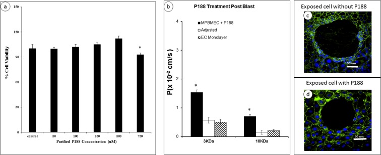
Dose dependent study of P188 on brain endothelial cell viability and modulated permeability coefficient. (a) Cell viability was measured after 24 h with MTT assay. Data represent mean ± SD from 6 independent experiments. *p < 0.05 when compared to the control (no P188 applied). (b) Changes in the permeability coefficients of the monolayer exposed to microcavitation and treated with P188 using 3 or 10 kDa Dextran molecules. *p < 0.05 compared to control (EC monolayer) from 6 independent experiments (n = 6). (c) Expansion of the crater if left untreated. Cells were exposed to microcavitation and incubated in serum-free media for 1 h and stained for ZO-1 (green) and nuclei (blue). (d) The crater expansion is prevented if the cells were treated with P188 (500 μm) for 6 hrs following microcavitation. Immunofluorescent images showed reconstitution of the tight junctions in response to P188.

