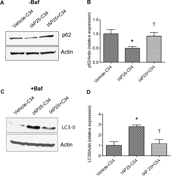
Figure 4.
IAP-induced autophagy is dependent on TLR4 pathway. RAW cells were pre-treated without (vehicle-C34, IAP-C34) or with TLR4 INC34 (IAP25 + C34) at 15 µM for 30 mins followed by addition of vehicle or IAP (A2356) (25U/ml) for 24 hours in the absence (A,B) or presence of Baf added for 3 hrs towards the end (C,D). (A) Western blotting showing the expression of p62. Actin was used a loading control. The gel was cut between 100 kDa and 25 kDa (probed for p62 ~62 kDa). Following detection of p62, the blots were stripped and re-probed for Actin. (B) Quantification of the blots using image J (mean ± SEM from three independent experiments). (C) Western blotting showing the expression of LC3-II in the samples treated with Baf. Actin was used a loading control. The gel was cut between 75 kDa (probed for actin ~37 kDa) and at 25 kDa (LC3-II at ~14 kDa) (D) Quantification of the blots using image J (mean ± SEM from three independent experiments). One way ANOVA and Dunnett’s Multiple Comparison Test was used to determine statistical significance. *P < 0.05 and Ϯ > 0.05, compared to vehicle control.

