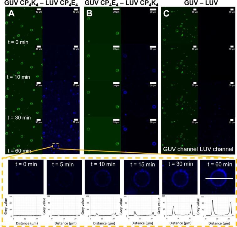
Figure 2.
Time-lapse micrographs of the lipid-mixing assay between lipopeptide-functionalized GUVs and lipopeptide-functionalized LUVs before (time = 0 minutes) and after (time = 10, 30 and 60 minutes) appearance of LUVs in the confocal volume. The GUVs are excited at 488 nm and fluorescence emission is detected between 500–550 nm (green), while LUVs are excited at 633 nm and emission is detected between 650–700 nm (blue). The lower panels show a selected GUV magnified at time = 0, 5, 10, 15, 30 and 60 minutes during the time-lapse lipid-mixing experiment and the corresponding fluorescence intensity evolution along the cross-section of the lipid bilayer is plotted in the graphs. (A) Lipid-mixing assay between CP4K4-functionalized GUVs and CP4E4-functionalized LUVs, (B) lipid-mixing assay between CP4E4-functionalized GUVs and CP4K4-functionalized LUVs, (C) lipid-mixing assay between non-functionalized GUVs and non-functionalized LUVs. Imaging was performed every minute for one hour using a Leica TCS SPE microscope.

