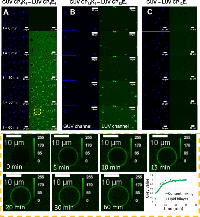
Figure 3.
Time-lapse micrographs of the content-mixing assay between lipopeptide-functionalized GUVs and lipopeptide-functionalized LUVs before (time = 0) and after (time = 5, 10, 30 and 60 minutes) appearance of LUVs in the confocal volume. GUVs are excited at 633 nm and fluorescence emission is detected between 650–700 nm (blue), while LUVs are excited at 488 nm and the emission is detected between 500–550 nm (green). The lower panels show one GUV magnified at time = 0, 5, 10, 15, 20, 30 and 60 minutes during the time-lapse content-mixing experiment and a normalized plot of the fluorescence evolution in the lumen and lipid bilayer of the GUV has been constructed from these images (lower right panel). (A) Content mixing assay between CP4K4-functionalized GUVs and CP4E4-functionalized LUVs, (B) content-mixing assay between CP12K4-functionalized GUVs and CP12E4-functionalized LUVs, (C) control content-mixing assay between non-functionalized GUVs and CP12E4-functionalized LUVs.

