Figure 1.
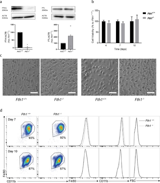
H-ferritin is not necessary for in vitro differentiation of bone marrow-derived macrophages. (a) Quantification of FTH1 and FTL by Western blot in protein extracts from Fth1+/+ (black) and Fth1−/− (grey) BMDM, at 10 days of culture. Quantification was made by densitometry analysis with ImageLabTM software of FTH1 or FTL bands normalized to β-actin (ACTB) in samples from 2–4 independent cultures and are represented as percentage relative to Fth1+/+ BMDM (t test *p < 0.05). (b) Cell viability of Fth1+/+ (black) and Fth1−/− (grey) BMDM was measured at 4, 7 and 10 days of culture, by resazurin reduction. The results represent the mean + SD of at least three independent cultures. The graphs depict the percentage of Fth1−/− viable cells relative to Fth1+/+ cultured in parallel. (c) Light microscopy images of BMDM at 7 (left panels) and 10 (right panels) days of culture. The cells were visualized and imaged in a Leica DMI6000 Time-lapse microscope. Images are representative of the cultures obtained from four animals per genotype. Scale bar: 50 μm. (d) Flow cytometry analysis of BMDM at 7 and 10 days of differentiation. Cells were stained for the myeloid markers F4/80 and CD11b. CD11b+ F4/80+ cells were considered completely differentiated macrophages. Left panels: flow cytometry plots gated for CD11b+ F4/80+ cells. Right panels: histogram plots of Fth1+/+ (solid black line) and Fth1−/− (dotted grey line) for F4/80, CD11b, and cell size (FSC).

