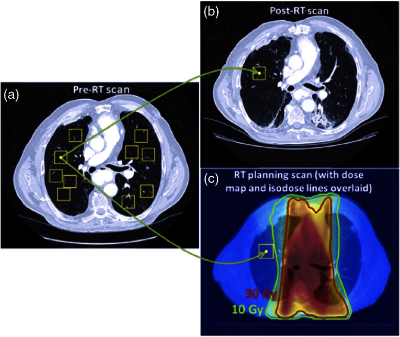
Fig. 2.
(a) ROIs are randomly placed in the lung volume of the pre-RT scans, and (b) the vector map obtained from deformable registration anatomically matches ROIs in the post-RT scan. (c) The vector map obtained from deformably registering the treatment planning scan is used to match ROIs in the pre-RT scan to the anatomical locations in the treatment planning dose map, assigning a dose distribution to each ROI. Only ROIs placed in high-dose regions () were used. Reprinted with permission from Ref. 22.

