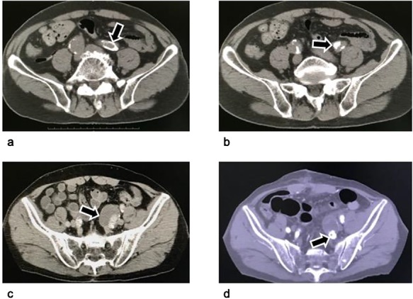
Figure 1.

Computed tomography of an isolated internal iliac artery aneurysm. (a–c): Images before the operation: An isolated internal iliac artery aneurysm on the left with a diameter of 36 mm can be seen. The left common iliac artery and the neck of the left internal iliac artery aneurysm were severely calcified (arrows). (d) Image four months postoperatively: The aneurysm sac has become smaller because its wall was resected as much as possible. The vascular plug in the internal iliac artery is visible distal to the aneurysm (arrow).
