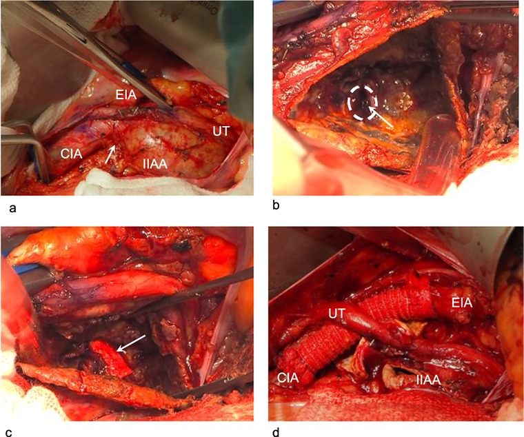
Figure 3.

Intraoperative findings during the open procedure as the second stage of hybrid intervention. (a) Dissection of the vessels proximal to the internal iliac artery aneurysm: We clamped the common iliac artery and external iliac artery for proximal control. The neck of the internal iliac artery aneurysm was very short (arrow). (b) Without surgical clamping of the distal branches, we opened the aneurysm. The origin of the distal internal iliac artery and the tip of the vascular plug obstructing it are visible. No bleeding into the internal iliac artery aneurysm can be seen. The first-stage embolization had completely controlled the distal blood flow. Arrow: the tip of the vascular plug. Circle: the orifice of the internal iliac artery distal to the aneurysm. (c) We ligated the origin of the distal internal iliac artery with a 4–0 pledgeted suture to avoid dislodgement of the vascular plug and delayed bleeding. Arrow: one of the two pledgets used in the ligation. (d) The common iliac artery could not be directly closed. We interposed the artery for proximal repair. The aneurysm wall was resected as much as possible.
CIA: common iliac artery, EIA: external iliac artery, IIAA: internal iliac artery aneurysm, UT: ureter.
