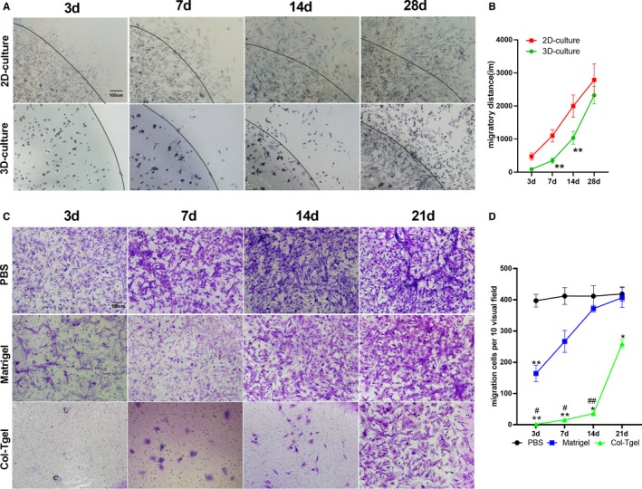
Figure 3.

Migration of adipose‐derived mesenchymal stem cells (ADSCs) in the transglutaminase cross‐linked gelatin (Col‐Tgel) scaffold. A, MTT staining of migrated adipose‐derived mesenchymal stem cells from gel spheres edge over time. B, The migratory distance of 2D‐PBS‐resuspended ADSCs and 3D‐Col‐Tgel encapsulated ADSCs (n=3 different samples per group at each time point). **P<0.01 vs 2D‐culture at corresponding time points. C, Transwell assay was performed at indicated time points after mixing ADSCs with PBS (upper), Matrigel (middle), and Col‐Tgel (Lower) compartment (n=3). ADSCs migrating through PBS, Matrigel, and Col‐Tgel were stained with crystal violet. D, The number of ADSCs migrated out of PBS, Matrigel, and the Col‐Tgel (n=3 different samples per group at each time point). Data were tested by 2‐way ANOVA without repeated measures, followed by a Bonferroni post hoc test. *P<0.05, **P<0.01 vs PBS, # P<0.05, ## P<0.01 vs Matrigel at corresponding time points.
