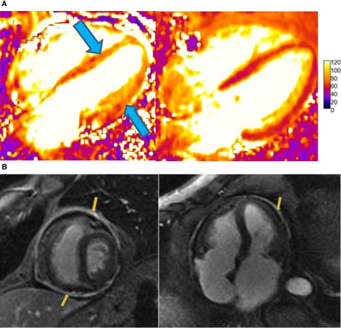
Figure 6.

A, (Left) Cardiac MRI T2 maps at diagnosis of myocarditis in a patient treated with pembrolizumab showing global hypokinesis with moderate systolic dysfunction (LVEF, 41%) and diffusely elevated T2 signal (arrows). (Right) T2 maps after withdrawal of pembrolizumab and 1‐month course of prednisone 1 mg/kg with resultant normalized systolic function (LVEF, 59%) and improved T2 signal. B, Cardiac magnetic resonance imaging scan late gadolinium enhancement (LGE) sequences showing thickened and enhanced pericardium in a patient with constrictive pericarditis following radiation therapy. Adapted with permission from Aldweib et al.123 Copyright © 2018, Eureka Science (FZC). LVEF, left ventricular ejection fraction.
