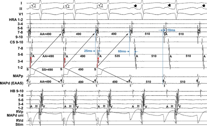
Figure 5.

Tracing during manifest entrainment by pacing from the distal coronary sinus (CS 1–2) in patient 13. The ECG leads I, II, and V1 and electrograms recorded from the high right atrium (HRA), coronary sinus (CS), mapping catheter (MAP), His bundle (HB) position, and right ventricle (RV) are shown. d indicates distal; EAAS, earliest atrial activation site; p, proximal; Stim, stimulation; uni, unipolar electrogram.
