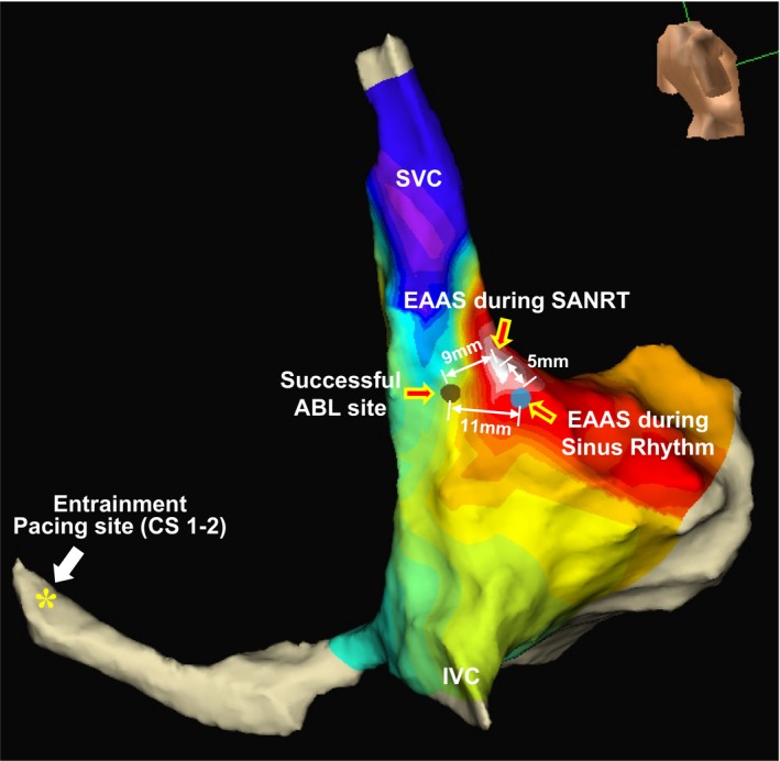
Figure 6.

Isochronal map during the tachycardia showing the relative locations of the earliest atrial activation site (EAAS) during the tachycardia, the EAAS during sinus rhythm, successful ablation (ABL) site, and entrainment pacing site (CS 1–2) in patient 13. CS indicates coronary sinus; IVC, inferior vena cava; SANRT, sinoatrial node reentrant tachycardia; SVC, superior vena cava.
