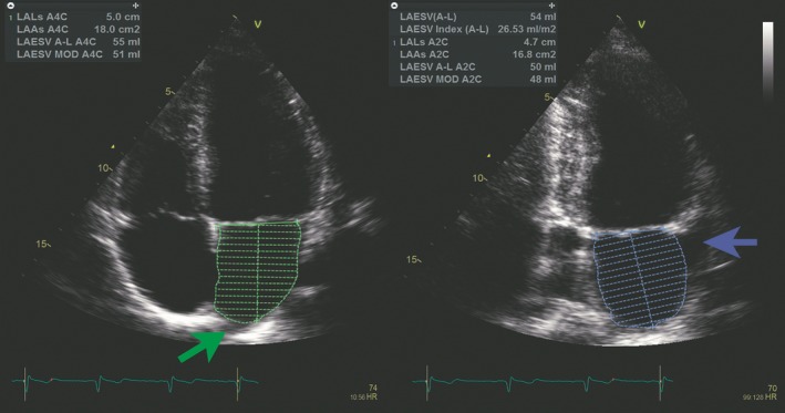
Figure 1.

Left atrial (LA) assessment. LA volume was measured from B‐mode recordings in apical 4‐chamber view (left) and 2‐chamber view (right). Tracing was done from one side at the mitral annular level following the endocardial border around the atrium and to the opposite site at the mitral annular level. The contour was closed at the mitral annulus with a straight line. The area of the atria in the specific view is annotated LAAs in the figure. Pulmonary veins (green arrow) and LA appendage (blue arrow) were excluded from the tracings. LA length was measured in both views, illustrated by the central line in the 2 tracings (annotated as LALs in the figure). LA volume was measured by the area‐length method (annotated as LAESV A‐L in the figure) and the summation of disks method (annotated as LAESV MOD in the figure).13
