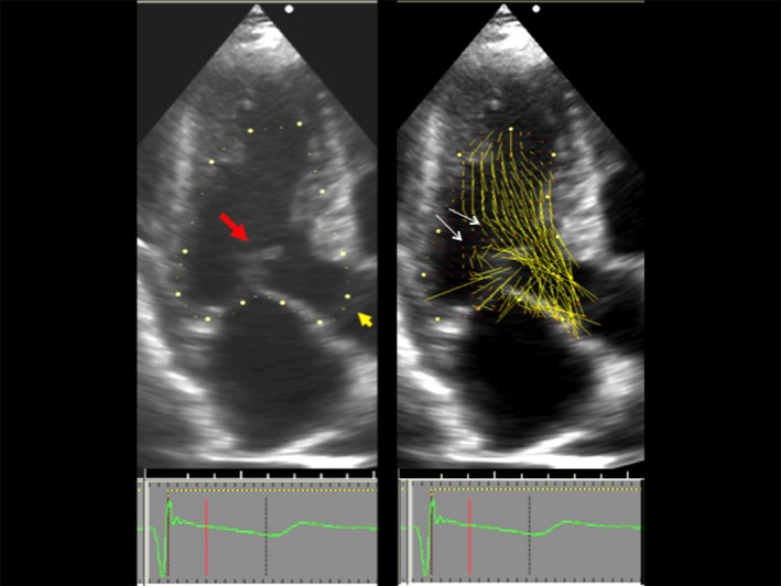
Figure 3.

Overlap between left ventricular outflow and the mitral valve. Left, Two‐dimensional echocardiogram, 3‐chamber view, showing the beginning of systolic anterior motion in systole. Red arrow points to mitral valve. Orange arrow points to the still closed aortic valve, also seen closed in the right panel. Right, Vector flow map of the same moment in systole. Local flow velocity is depicted as yellow lines proportional to, and in the direction of, local velocity, indicated by red head of the vector. The overlap between the ejection flow in the outflow tract and the mitral valve is seen. The septal bulge displaces ejection flow so that it comes from a more posterior direction. Note that vector flow impacts the posterior surface of the mitral leaflets with high angle of attack (white arrows).3
