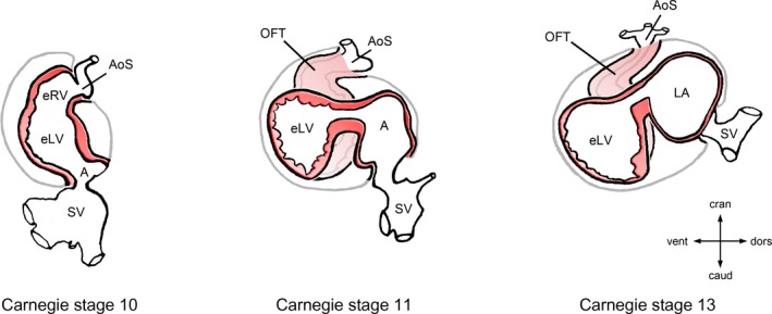
Figure 4.

Ascent of the atria during looping morphogenesis of the embryonic heart. This diagram depicts the progressive changes of the topographical relationship between the developing atrial and ventricular chambers of the human embryonic heart during the fourth and fifth week of development (gestational weeks 6 and 7). Hearts are shown in left lateral views. During the initial phase of heart looping (C‐looping; Carnegie stage 10), the embryonic atrium is caudal and dorsal to the embryonic ventricles. During S‐looping (Carnegie stages 11–13), the future atrial chambers are shifted from their original caudal position, first toward a dorsal position (Carnegie stage 11) and, later, toward their definitive position dorsal and cranial to the embryonic ventricles (Carnegie stage 13). The ascent of the atria occurs in all terrestrial vertebrates and sets the scene for establishment of the 180° U turn between the atrioventricular and semilunar valves found in the mature heart of birds and mammals. A indicates embryonic atrium; AoS, aortic sac; caud, caudal; cran, cranial; dors, dorsal; eLV, embryonic left ventricle; eRV, embryonic right ventricle; LA, left half of the common embryonic atrium; OFT, outflow tract; SV, sinus venosus; vent, ventral.
