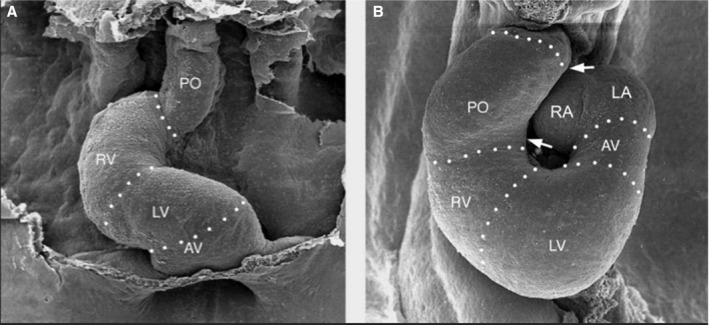
Figure 5.

Ascent of the atria. Scanning electron micrographs, viewed from front, show the heart loops of chick embryos at the end of C‐looping (left) and later after early S‐looping (right). Note that at the end of C‐looping, the venous pole of the heart is in a primitive position caudal to the embryonic ventricles. During early S‐looping, the future atrial chambers are shifted from their original caudal position toward their definitive position cranial to the embryonic ventricles. AV indicates atrioventricular canal; LA, left half of the common atrium; LV, embryonic left ventricle; PO, proximal part of outflow tract; RA, right half of the common atrium; RV, embryonic right ventricle. Reprinted from Männer30 with permission. Copyright ©2008, John Wiley and Sons.
