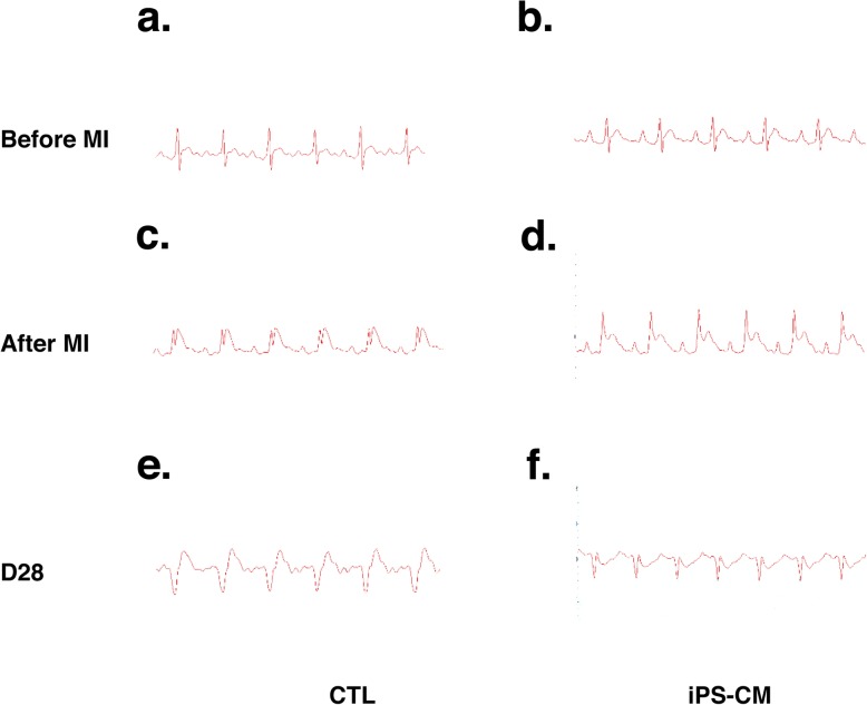
Fig. 2.
Electrocardiogram (ECG) for rats during establishment of the myocardial infarction (MI) model and 28 days after iPSC-CM therapy. Representative traces from rat ECG recordings are shown (a, b). Readings are normal at baseline before left anterior descending (LAD) coronary artery ligation in the CTL group and iPS-CM group. c, d ST segment elevation in every group after LAD coronary artery ligation, which indicated that the MI model was successful. e, f No ventricular arrhythmias occurred 28 days after iPSC-CM transplantation. CTL control, iPSC-CMs induced pluripotent stem cell-derived cardiomyocytes

