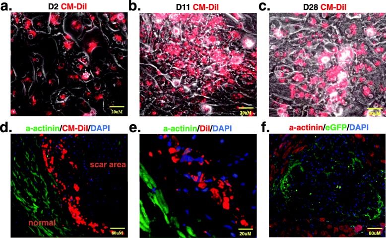
Fig. 5.
Histological evaluation confirms iPSC-CM homing to the infarcted heart. a–c The grafted cells were labeled with the fluorescent cell tracer Vybrant™ CM-DiI Cell-Labeling Solution (Molecular Probes, Invitrogen) and cultured in vitro for 2 days (a), 11 days (b), and 28 days (c). d, e Immunofluorescent staining shows alpha-actinin (green) in rat heart tissue; the grafted cells were prelabeled with Vybrant- CM-DiI (Red) and detected under a microscope 7 days after transplantation. f iPSC-CMs were detected by eGFP immunofluorescent staining 4 weeks after transplantation. iPSC-CMs induced pluripotent stem cell-derived cardiomyocytes, and eGFP enhanced green fluorescent protein

