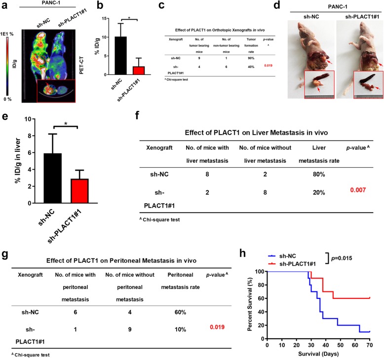
Fig. 4.
PLACT1 facilitates the tumorigenesis and metastasis of PDAC in an orthotopic xenograft model. a-b Representative PET-CT images (a) and histogram analysis (b) of 18FDG accumulation in pancreas in orthotopic xenografts after orthotopically injections with indicated PANC-1 cells (n = 10). The 18FDG concentrations in orthotopic tumor were normalized to %ID/g. c The tumor formation rate of orthotopic xenograft was calculated for indicated group (n = 10). d Representative images of orthotopic tumors after orthotopically injections with indicated PANC-1 cells. e Histogram analysis of 18FDG accumulation in liver in orthotopic xenografts after orthotopically injections with indicated PANC-1 cells (n = 10). f-g The liver metastasis (f) and peritoneal metastasis (g) rate of orthotopic xenograft was calculated for indicated group (n = 10). h Survival analysis for orthotopic tumor bearing mice in indicated group (n = 10). Statistical significance was assessed using two-tailed t-tests and ANOVA followed by Dunnett’s tests for multiple comparison. Error bars represent standard deviations of three independent experiments. *p < 0.05 and **p < 0.01

