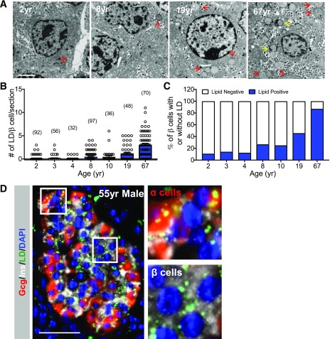
Figure 1.
LD accumulation in human islet β-cells increases with age. A: Representative EM images of healthy human islet β-cells from a 2-, 8-, 19-, and 67-year-old donor, which illustrates that very few LDs (red arrows) are found in juvenile donors compared with adult. The yellow arrows depict lipofuscins. Scale bar = 2 μm. yr, years. B: The number of lipid granules (including lipofuscins) per donor β-cell, with the quantity of β-cells counted in parenthesis. C: Percentage of β-cells containing lipid granules in human donors of different ages. D: Representative immunofluorescent image showing LD accumulation in healthy adult islet α- and β-cells from a 55-year-old (55yr) donor. Staining was performed to detect insulin (Ins) (white), glucagon (Gcg) (red), LDs (BODIPY [green]), and nuclei (DAPI [blue]). Scale bar = 50 μm. Right: magnified view of LD build-up in α- and β-cells within the marked area in the left panel. Error bars indicate SEM.

