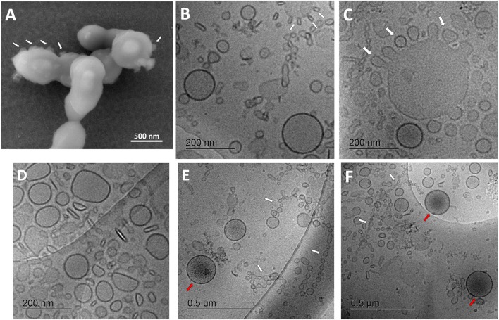
Figure 2.
Characterization of streptococcal membrane vesicles (MVs) using electron microscopy. (A) Scanning electron micrograph (SEM) of Streptococcus pneumoniae reference strain R6 cells, during shedding of vesicles from their surface. (B–F) Cryogenic transmission electron micrographs (cryo-TEM) of isolated pneumococcal MVs, showing heterogeneous morphology, tiny vesicles budding from large ones (white arrows), chain-like structures (white arrows), and some vesicles with darker content (red arrows).

