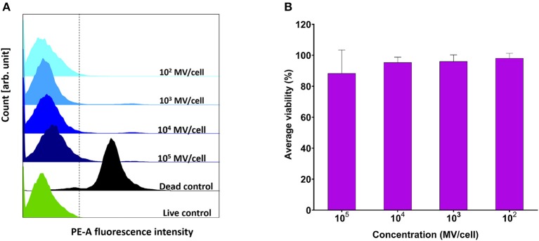
Figure 4.
Viability assessment of dendritic cells (DC2.4) after exposure to pneumococcal membrane vesicles (MVs) by flow cytometry. (A) Overlay of recorded fluorescence intensity of phycoerythrin (PE-A) channel, after exposure of DC2.4 cells to various concentrations of MVs, in addition to live and dead cell controls. (B) Calculated percentage viability of DC2.4 cells with respect to cells treated with PBS (live cells control) and 4% PFA (dead cells control), after exposure to streptococcal MVs. The dashed line separates PE-A channel negative and positive cells. n = 3, mean ± SD.

