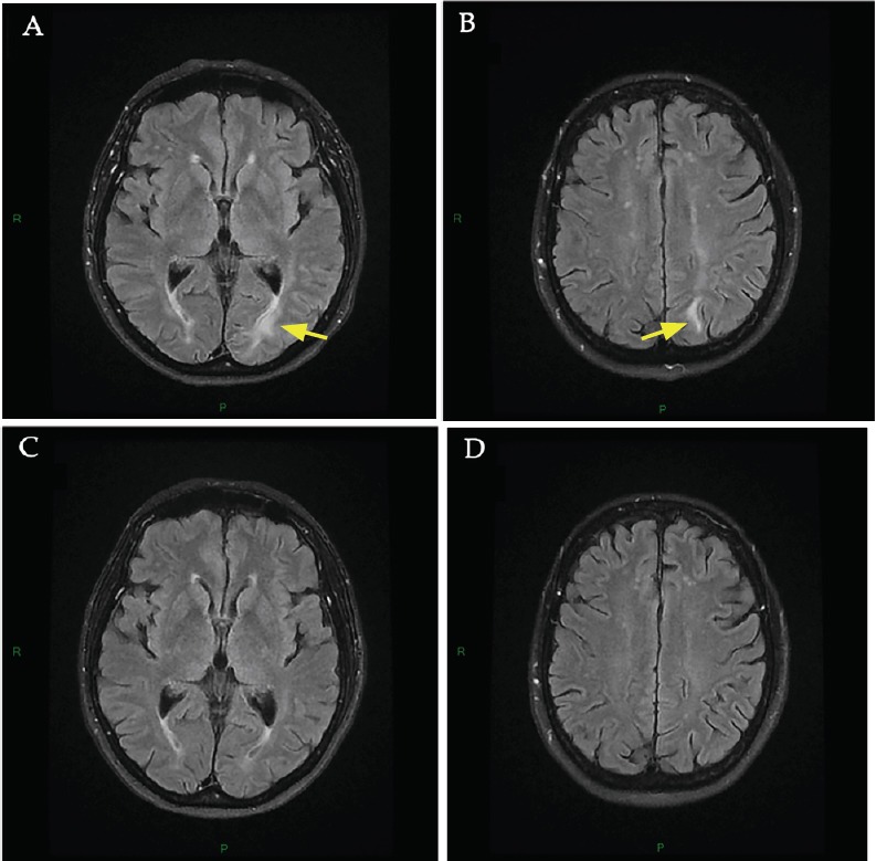
Figure 2.

Brain MRI at onset of neurological disturbances (day 16 after pazopanib initiation) and 3-month follow-up.
(A, B) Brain MRI at onset of neurological disturbances. MRI at onset of PRES showed hyperintensities in the left occipital (A, arrow) and the left parietal (B, arrow) regions involving the white matter in T2-FLAIR sequence. No diffusion abnormalities were found in the diffusion weighted imaging sequence and apparent diffusion coefficient was increased. These lesions were consistent with a vasogenic edema of PRES. Major differential diagnoses were excluded including posterior reversible vasoconstriction syndrome (time-of-flight MRI), cerebral bleeding (T2* MRI) and stroke. (C, D) 3-month follow-up brain MRI. New brain MRI 3 months later showing complete resolution of the lesions of PRES in the left occipital (C) and the left parietal regions (D).
