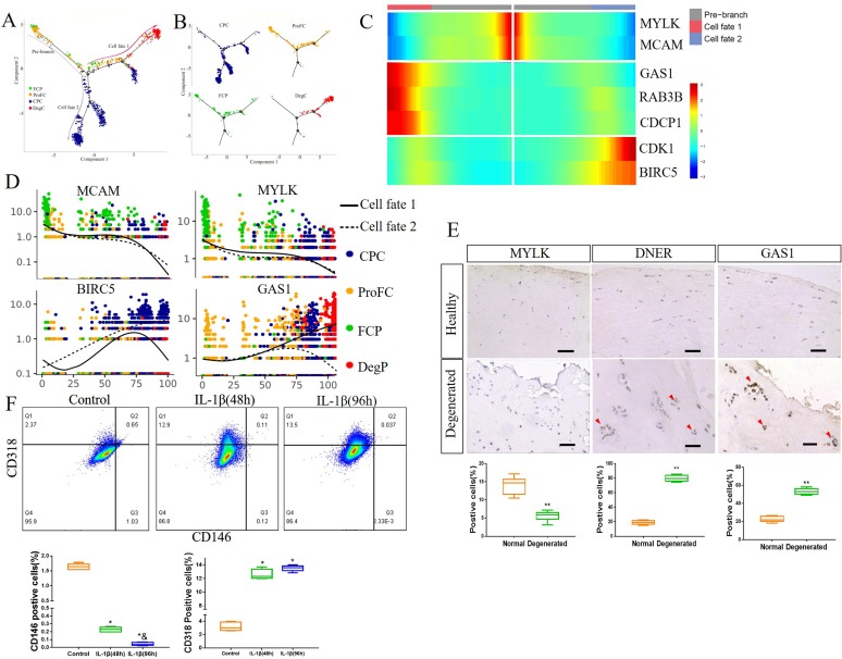
Figure 5.
Identification of degenerated meniscus progenitor cells (DegP) as a key element for meniscus degeneration. (A, B) Monocle pseudotime trajectory showing the progression of FCP, ProFC, CPC and DegP. (C) From the centre to the left of the heatmap, the kinetic curve from the root along the trajectory to fate 1. Starting from the right, the curve from the root to fate 2. FCP markers MYLK and MCAM, DegP markers GAS1, Rab3B and CDCP1 and CPC markers CDK1 and BIRC5 expressed from the root to each branch. (D) Pseudotime kinetics of indicated genes from the root of the trajectory to fate 1 (solid line) and the cells up to fate 2 (dashed line). (E) Representative IHC staining of MYLK, GAS1 and DNER in healthy human meniscus and degenerated meniscus, and quantification of positive cells. Scale bar, 50 µm. n=6, **p<0.01. (F) Healthy human meniscus cells were treated with 5 ng/mL IL-1β for 48 hours or 96 hours. Phosphate buffer saline (PBS) was used as a negative control. CD146 and CD318 expression was determined by flow cytometry. n≥5, * versus control, p<0.05; & versus IL-1β (48 hours), p<0.05. CPC, cartilage progenitor cells; DeP, degenerated meniscus progenitor cell; FCP, fibrochondrocyte progenitors; ProFC, proliferate fibrochondrocytes.

