Figure 6.
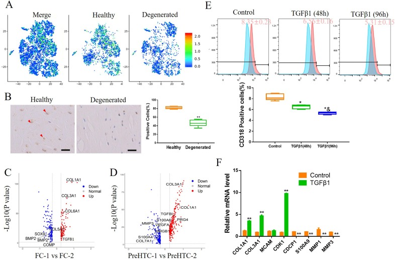
Activation of TGFβ signalling pathway attenuates the increase in CD318+ cells in degenerated meniscus. (A) The expression of TGFβ1 on merged and split t-distributed stochastic neighbourembedding map. (B) IHC staining of TGFβ1 on human healthy meniscus and degenerated meniscus. n=6, **p<0.01. (C) Volcano plot comparing the gene expression between FC-1 and FC-2. Each plot represents one gene. (D) Volcano plot comparing the gene expression between PreHTC-1 and PreHTC-2. Each plot represents one gene. (E) Human degenerated meniscus cells were treated with 5 ng/mL TGFβ1 for 48 hours or 96 hours. PBS was used as a negative control. CD318 expression was determined by flow cytometry (n≥5). * vs control, p<0.05; & vs TGFβ1 (48 hours), p<0.05. (F) Human degenerated meniscus cells were treated with 5 ng/mL TGFβ1 or PBS as negative control. The expression of indicated marker genes were detected by qRT-PCR. n=3, **p<0.01.

