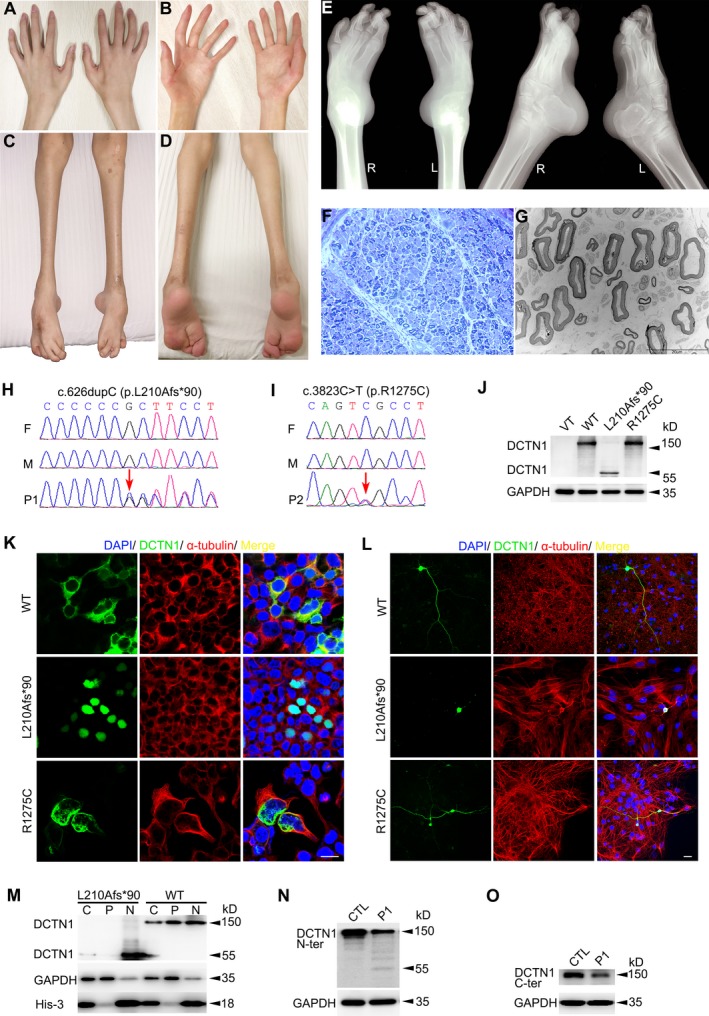
Figure 1.

Clinical, pathological, genetic, and functional characterizations. (A–D) Hands (A and B) and lower limbs (C and D) of Patient 1. (E) X‐ray photograph of lower limbs of Patient 1 showing bilateral talipes equinovarus. (F and G) Peripheral nerve pathology of the Patient 1 showed mild density decrease of large myelinated fibers by toluidine blue staining (F) and under electronic microscopy (G). (H) Sequence chromatograms of DCTN1 gene of Patient 1’s family. It displays one de novo insertion mutation of c.626dupC (arrow) in the proband. F = father, M = mother, P1 = patient 1. (I) Sequence chromatograms of DCTN1 gene of Patient 2’s family. It displays one de novo missense mutation of c.3823C>T (arrow) in the proband. (J) Western blotting showed the expressions of DCTN1‐Mut (L210Afs*90) were significantly lower than healthy control. Western blotting showed the DCTN1‐WT and R1275C with molecular weight of 150 kDa. DCTN1‐L210Afs*90 with relative smaller molecular weight (∼55 kDa) was detected. (K) DCTN1‐WT/Mut‐EGFP transfected HEK 293T cells showing the presence of WT in cytoplasmic distribution colocalizing with α‐tubulin but L210Afs*90 is expressed in nuclear and R1275C forms punctate aggregates. The scale bar represents 20 μm. (L) Immunofluorescence of DCTN1‐WT/Mut in primary mouse cortex neuron showing WT and R1275C is expressed in both body and axon, but L210Afs*90 is only expressed in the body. The scale bar represents 20 μm. (M) Western blotting of separated whole cell/nuclear/cytoplasmic components showed the L210Afs*90 is mainly expressed in nuclear. GAPDH and His‐3 are set as housekeeping proteins. C = cell, N = nuclear, P = cytoplasmic. (N–O) Western blotting showed a strong signal at ~150 kDa of skin fibroblasts from control samples. There was relative weak signal detected in the Patient 1’s protein using either N‐terminus (N) or C‐terminus antibody (O). A signal at relative smaller molecular weight (~55kDa) was detected in the patient but not in control using N‐ terminus antibody (N).
