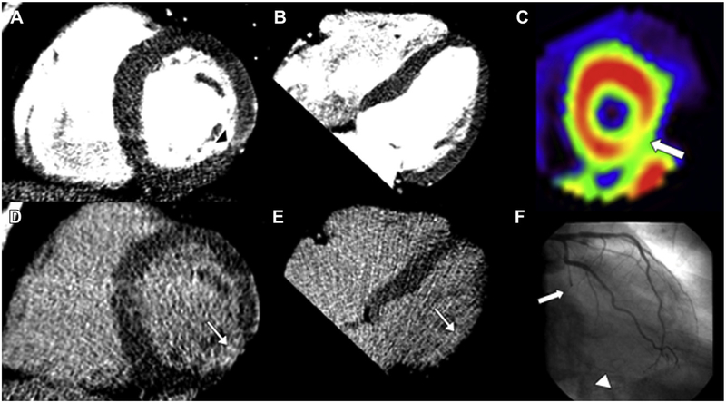
Figure 3.
Circumflex infarct. Cardiac CT images (A, B, D, and E), SPECT (C), and invasive angiography (F) of a 63-year-old man with a history of dyslipidemia, myocardial infarction, and percutaneous coronary intervention (left circumflex, left anterior descending, diagonal branch). Regional myocardial thinning in the lateral wall (black arrowhead) on the rest images (A and B) and abnormal late contrast enhancement (white arrow) on the delayed images (D and E) indicate the presence of infarction in the circumflex territory (inferolateral wall). The SPECT image of the short axis of the left ventricle shows moderate ischemia in the inferolateral wall (C; white arrow). The angiogram shows occlusion of the mid circumflex (F; white arrow) and filling of distal collaterals (arrowhead).

