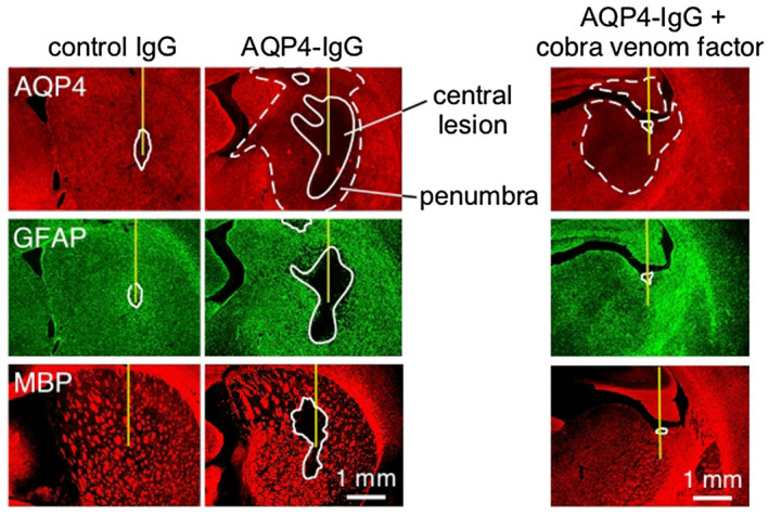
Figure 3.

NMO pathology produced in rat brain following intracerebral injection of AQP4‐IgG. AQP4, GFAP and MPB immunofluorescence at 5 days after injection of control (non‐NMO) IgG or AQP4‐IgG in rats. Solid line shows central area of fluorescence loss and dashed line denotes penumbra region seen for AQP4. Where indicated (right), rats were pretreated with cobra venom factor to inactive complement. Adapted from Ref. 2.
