Figure 5.

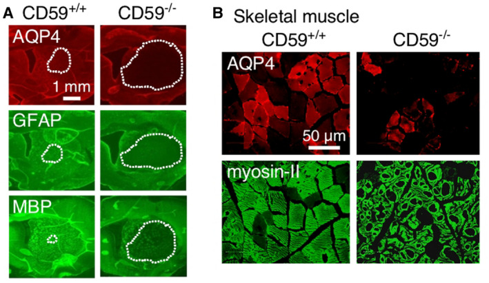
CD59 knockout in rats greatly amplifies NMO pathology following passive transfer of AQP4‐IgG. A. AQP4, GFAP and MBP immunofluorescence of rat brain at 7 days after intracerebral injection of AQP4‐IgG in wild‐type (CD59+/+) and knockout (CD59−/−) rats. Dotted line denotes areas of loss of fluorescence. Adapted from Ref. 67. B. AQP4 and myosin‐II immunofluorescence at 24 h after intraperitoneal injection of AQP4‐IgG in CD59+/+ and CD59−/− rats. Adapted from Ref. 66.
