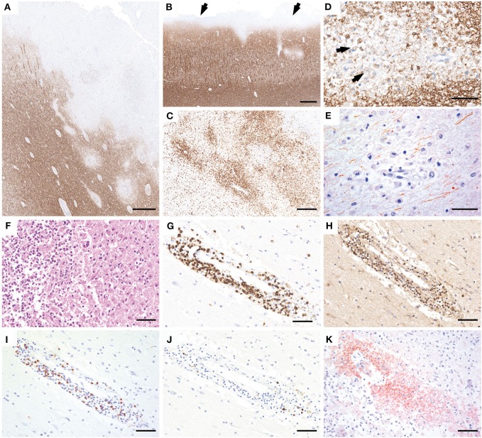
Figure 2.
Neuropathology of brain autopsy. Histopathology of the brain autopsy reveals numerous, predominantly cortical demyelinating lesions that expand into the subcortical white matter (A; MOG) or form a subpial band of demyelination (B, arrows). The lesion borders show numerous activated microglia and macrophages (C, HLADR). The plaques are characterized by partly confluent, partly perivenous areas of demyelination, with MOG-positive myelin degradation products within the macrophage cytoplasms (D, arrows). Axons are relatively better preserved but the density is clearly reduced compared to the peri-plaque white matter (E, SMI31). Some lesions show superimposed ischemic damage with tissue necrosis with pronounced infiltration of neutrophilic granulocytes (F, H&E). The inflammatory infiltrates are mainly composed of CD3 (G) and CD4-positive T cells (H), less CD8-positive T cells (I), and few perivascular CD79a-positive B cells (J). Within the lesions, profound perivenous deposition of activated complement complex is visible (K, C9neo antigen). Scale bars: (A,B) 600 μm; (C) 300 μm; (D,E) 50 μm; (F–K) 60 μm.

