
Figure 1.
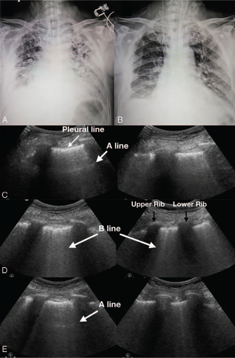
Imaging studies of chest x-ray and lung ultrasound. Chest x-ray showed bilateral diffuse and heterogeneous opacities on the first day of ICU admission (Panel A). Opacities diminished markedly within 1 week (Panel B) with bilateral A-line on the anterior chest (Panel C, left panel) and multiple B-line on the posterolateral chest wall (Panel C, right panel) evaluated by lung ultrasound examination. One day later, examination showed a marked increase of B lines bilaterally and a disappearance of A lines on the anterior chest (Panel D, left panel: upper point, right panel: lower point). After 24 hours of treatment, a repeated lung ultrasound revealed a marked decreased of B lines and A lines reappeared on the anterior chest. (Panel E, left panel: upper point, right panel: lower point). (The A line: the horizontal lines arising from the pleural line are separated by regular intervals that are equal to the distance between the skin and the pleural line. The A-line indicates air. The B line: multiple vertical comet-tail artifacts arising from the pleural line are spreading to the edge of the screen without fading and moving with lung sliding. It reflects the coexistence of elements with a major acoustic impedance gradient, such as fluid and air. Several B lines indicate increasing pulmonary fluid.). ICU = intensive care unit.