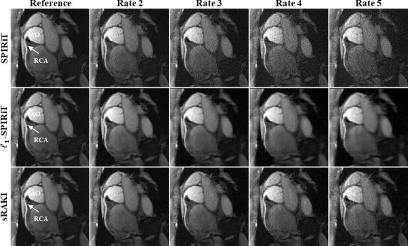
Fig 2. Reformatted right coronary artery (RCA) images from a 3D targeted coronary MRI dataset.

The data were retrospectively undersampled at rates 2, 3, 4, and 5 in the ky—kz plane and then reconstructed using SPIRiT, l1-SPIRiT and sRAKI (top, middle and bottom rows). Fully-sampled images are also displayed in the first column as a reference for comparison. sRAKI is visually more robust to noise amplification and blurring artifacts at high acceleration rates compared to SPIRiT and l1-SPIRiT, respectively. (RCA: right coronary artery; AO: Aortic Root).
