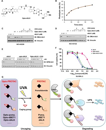
Fig. 4. Light controls the effects of opto-dALK in mediating the degradation of the ALK fusion protein.

(A) A schematic illustration of the chemical structure of the engineered opto-dALK. (B) Time course uncaging of opto-dALK by UVA irradiation in vitro. Opto-dBET1 (1 mM) was irradiated with UVA (365 nm) for indicated time and then subjected to the UV-Vis absorption analysis. (C to E). UVA irradiation activates opto-dALK to promote the degradation of EML-ALK fusion proteins in cells. IB analysis of WCL derived from NCI-H2228 (C) or NCI-3122 (D) NSCLC cells or SU-DHL-1 cells (E) treated with BET1 versus opto-dBET1 at indicated dose with or without UVA irradiation (365 nm) for 5 or 15 min. (F) UVA irradiation–activated opto-dALK inhibits SU-DHL-1 cell proliferation in a dose-dependent manner. SU-DHL-1 cells were treated by dALK versus opto-dALK with or without UVA irradiation (365 nm) for 15 min and then subjected to CCK-8 cell viability assay. (G) A schematic diagram showing that working model of opto-pomalidomide in degrading POI in a UVA-dependent manner.
