
Fig. 5. Optical control of BRD2–4 levels.
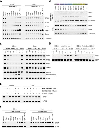
(A) Immunoblot analysis after treatment of RS4;11 cells with PHOTAC-I-3 for 4 hours at different concentrations. Cells were either irradiated with 100-ms pulses of 390-nm light every 10 s (left) or kept in the dark (right). MLN (MLN4924) was added as an additional control. (B) Time course of BRD2–4 degradation, c-MYC levels, and PARP1 cleavage assayed by immunoblotting. RS4;11 cells were treated with PHOTAC-I-3 (1 μM) and collected at the indicated time points. PHOTAC-I-3 has no effect on BRD2–4 levels in the dark over several hours. (C) Immunoblot of a rescue experiment demonstrating the reversibility of degradation promoted by PHOTAC-I-3 through thermal relaxation (left) or optical inactivation by 525-nm pulsed irradiation (right, 100 ms every 10 s). (D) Color dosing: Wavelength dependence of BRD2/4 degradation promoted by 300 nM PHOTAC-I-3. (E) Immunoblot analysis after treatment of RS4;11 cells with PHOTAC-I-3 and combinations with lenalidomide or (+)-JQ1 for 4 hours to confirm a CRBN-based mechanism. Cells were either irradiated with 100-ms pulses of 390-nm light every 10 s (left) or kept in the dark (right). (F) Optical degradation of BRD4 with the thalidomide derivative PHOTAC-I-10. Immunoblot analysis of RS4;11 cells after treatment with PHOTAC-I-10 for 4 hours at different concentrations, which were either irradiated with 100-ms pulses of 390-nm light every 10 s (left) or kept in the dark (right).