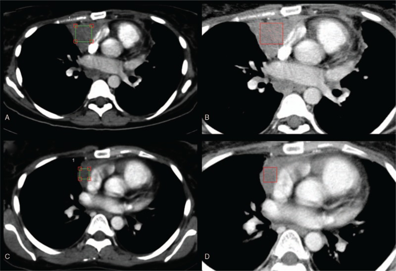
Figure 1.

Contrast-enhanced computed tomography texture analysis (CTTA) of a Hodgkin lymphoma patient with a lymphoma mass in the anterior mediastinum before (upper row) and after (lower row) a standardized chemotherapy protocol. For quantification of tissue texture, cubiform volume of interests were drawn inside the lymphoma mass excluding adjacent vessels (A and C). Color-coded CTTA maps display the mean intensity of the lymphoma mass before (B) and after (D) chemotherapy using a fine filter.
