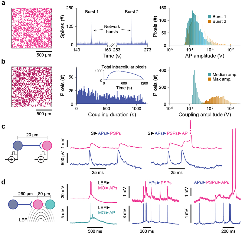
Fig. 3 |. Network-wide intracellular measurements of dissociated rat neurons in the pCC configuration.

a, Intracellular recordings across the array using Ie = -1.1 nA show synchronized firings: large network bursts involving 1,837 pixels (left, Burst 1) and 1,882 pixels (Burst 2), show correlated spikes (middle, histogram bins of 10 ms) at 153 s and 263 s of the experiment, respectively. The AP spike amplitudes are extracted from both events (right), with an array median of 172 μV and 229 μV, respectively. b, Another array-wide intracellular recording, with continual stimulation and a total of 1,728 pixels (left) intracellularly coupled throughout the 19-minute recording. The coupling duration (middle, median of 495 s) and AP spike amplitude (right) are characterized. The total number of simultaneously intracellularly coupled pixels as a function of time is plotted in the middle inset, with a maximum of 982 pixels being simultaneously intracellularly coupled at time 396 s. c, Array-wide stimulation increases the overall synaptic network activity: stimulated APs of the presynaptic neuron (blue: S►APs) induce excitatory PSPs in the postsynaptic neuron (magenta, APs►PSPs). When the PSPs are close enough to summate and exceed threshold, an AP fires (PSPs►APs). d, Spontaneous (non-stimulated) membrane potentials of the middle neuron (magenta) can be related to the left neuron via their synaptic connection (blue) and the local electrochemical field (LFF) that also excites the right neuron (green). APs from the blue neuron induce excitatory PSPs that in turn induce APs. The LEF induces membrane oscillations (MOs); large MOs cause the neurons (magenta and green) to exceed their thresholds to fire APs (MO►APs).
