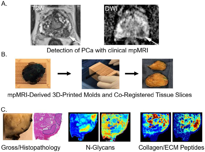
Figure 1. An integrated three dimensional multimodality work flow to evaluate new glycomic and extracellular matrix biomarker candidates for prostate cancer.
A. Representative pre-prostatectomy MRI images for T2W (T2 Weighted Image) and DWI (Diffusion-weighted image). B. A representative 3D-mold from mpMRI used for preparing prostatectomy tissue co-registered with mpMRI coordinates. C. Gross pathology and standard H&E stained tissue section, with two representative MALDI-IMS images of tumor and stroma localized N-glycans and collagenase derived ECM peptides in the same tissue slice.

