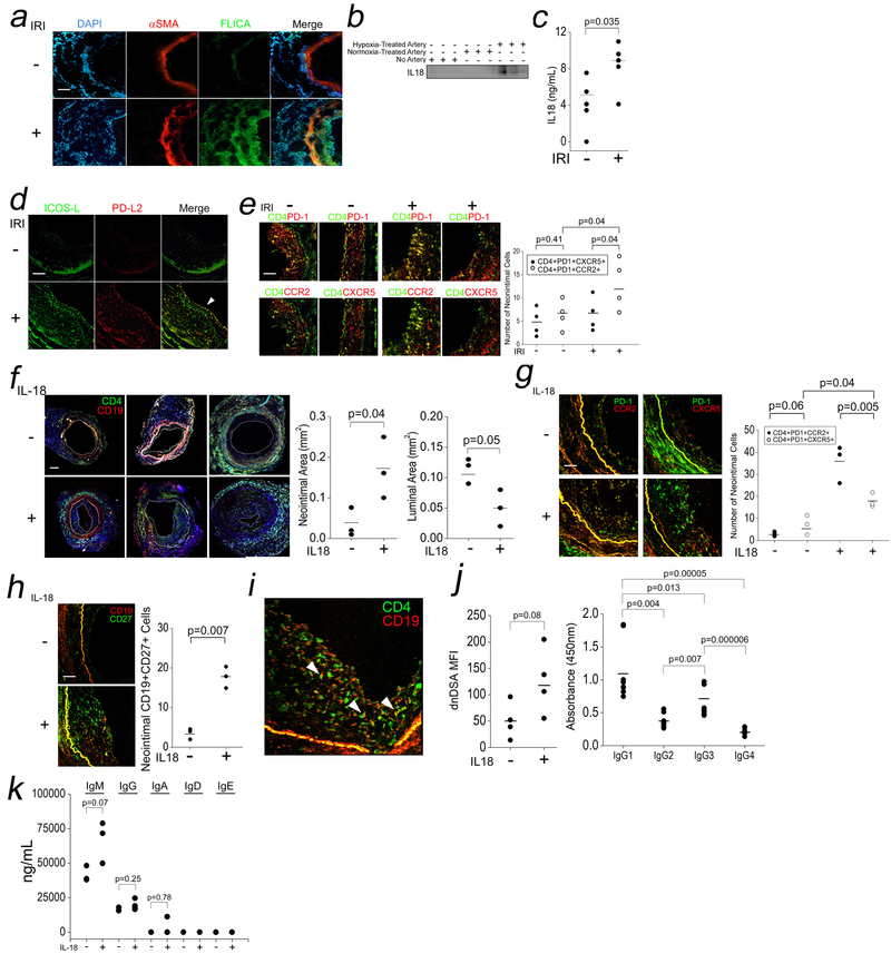
Fig 5. IRI-Induced Inflammasomes in EC and IL-18-Mediated TPH Cell Expansion In Vivo.
Human coronary artery grafts were subjected to ex vivo hypoxia and surgically implanted into descending aortae of SCID/bg mice for 24h prior to analysis by I.F. for FLICA (a, scale bar: 200μm) and sera were analyzed by Western blot IL-18 (b) and ELISA (c). n=3–5 for the above experiments. Grafts were analyzed for ICOS-L and PD-L2 (d, 250μm) and TPH and TFH cell infiltrates (e, scale bar: 250μm). Hosts bearing normoxia-treated human arteries were injected i.p. with vehicle or IL-18 (10μg/dy) for 14 days. Neointimal and luminal areas were calculated (f, scale bar: 400μm), and neointimal TPH cells and TFH cells were quantified (g, scale bar: 250μm) along with CD19+CD27+ B cells (h). CD4+:CD19+ cell “conjugates” were visualized in neointimal tissues (i). Sera was tested for dnDSA Ab titers (j, left), dnDSA IgG subclasses (j, right), and Ig isotypes (k). Student’s t-test was used for Fig 5c, 5f, 5h, and 5j, left. One-way ANOVA followed by Tukey’s pairwise comparison was used for Fig 5j, right, and 5k. Two-way ANOVA followed by Tukey’s pairwise comparison was used for Fig 5e and 5g.

