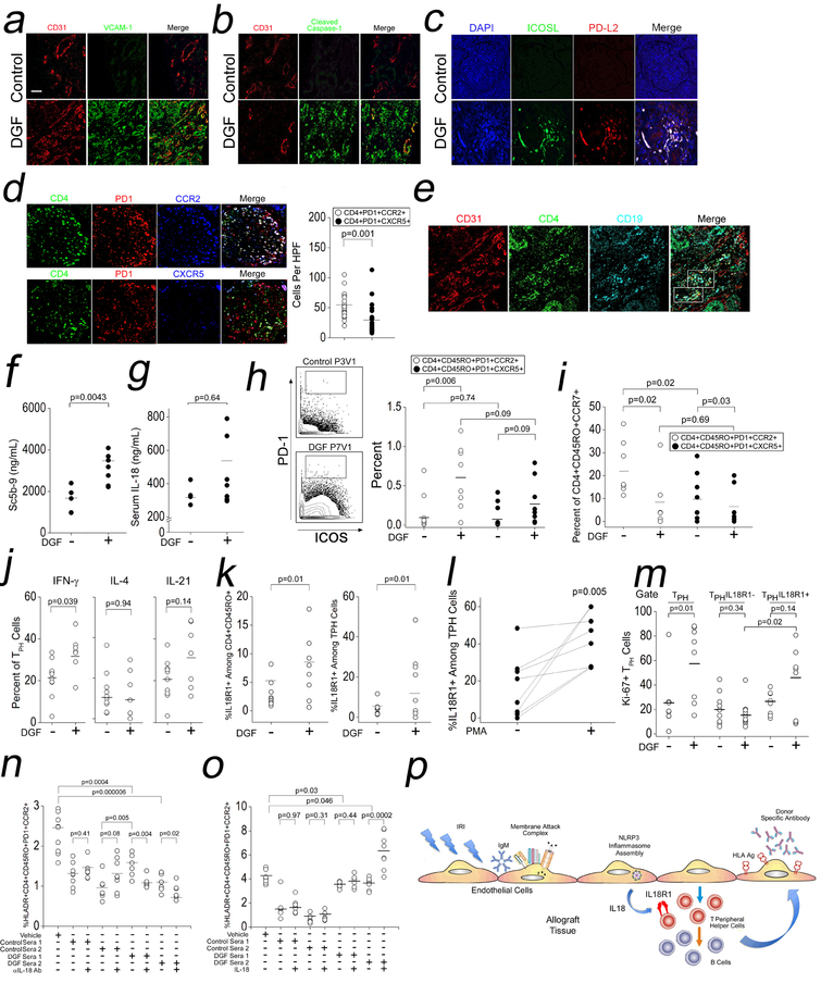
Fig 6. IL-18-Dependent Expansion of IL-18R1+ TPH Cells in DGF Patients.
Archived biopsies from patients with DGF who developed CABMR were analyzed by I.F. (a-e). Prospectively collected sera from control or DGF renal transplant patients were assessed for complement activation (f) and IL-18 (g). PBMCs from control or DGF renal transplant patients were analyzed by CyTOF (h-m). In (j) and (l), PBMCs were stimulated for 4h with PMA/ionomycin prior to CyTOF analysis. Non-autologous Tmem were stimulated with αCD3/CD28 for 24hr in the presence of 10% v/v autologous sera from DGF or control patients in the presence αIL-18 Ab (n) or exogenous IL-18 (o). Proposed model connecting IRI with CABMR (p). Scale bars: 300μm. Student’s t-test was used for Fig 6d, 6f, 6g, 6j, 6k, and 6l. Two-way ANOVA followed by Tukey’s pairwise comparison was used for Fig 6h, 6i, 6m, 6n, and 6o.

