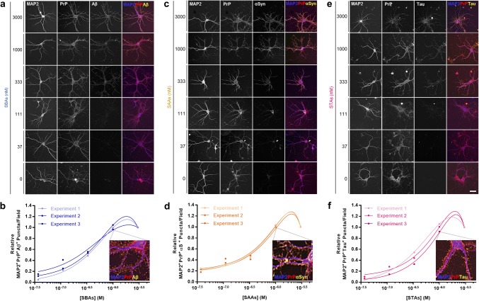
Fig. 3.
SBAs, SAAs and STAs bind to primary neurons in a dose-dependent and saturable manner. a, c, e Soluble protein aggregates were added to primary mouse neurons (MPNs) and binding assessed using serial-permeabilized immunocytochemistry. Representative images SBAs (a), SAAs (c) and STAs (e) are shown. Staining for MAP2, PrP and bound proteins are shown in blue, red and yellow, respectively. b, d, f Relative dose–response binding of SBAs (a), SAAs (c) and STAs (e) to MPNs was quantified using a custom FIJI macro that identified soluble protein aggregate puncta that were at least 50% PrP colocalized. Relative values were determined by normalizing binding signals to those obtained with the highest protein concentration analyzed (3 µM). Inset images depict enlarged triple-colocalization images for neurons treated with 1 μM SPAs (from a, c and e). Scale bar in a, c and e = 50 µm, and data in b, d and f represent three independent experiments with 60 images analyzed per experiment per dose

