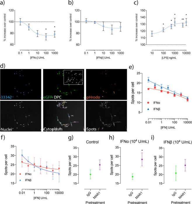
Figure 1.
Treatment with IFN inhibits phagocytosis in both BV-2 and primary CX3CR1eGFP/+ primary microglia, with IFNα inhibition reversible with an anti-IFNAR1 neutralising antibody. Phagocytosis was determined using pHrodo E. coli fluorescent particles and is represented as % increases over untreated samples following varying concentrations of (a) IFNα, (b) IFNβ and (c) LPS (*p < 0.05, **p < 0.01, 1-way ANOVA, Dunnett’s multiple comparisons test, n = 5–6). (d) Workflow of imaging analysis for pHrodo E.coli treatments for both BV-2 and CX3CR1eGFP/+ primary microglia. Analysis of spots per cell following varying concentrations of IFN in (e) BV-2 cells and (f) primary CX3CR1eGFP/+ microglia. Analysis of spots per cell following IgG and MAR1 pre-treatment after which cells were exposed to (g) control (h) 104 U IFNα and (i) 104 U IFNβ (*p < 0.05, Students unpaired t-test, n = 5–6). Data is expressed as mean ± SEM.

