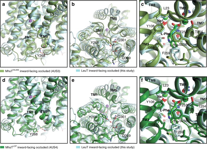
Fig. 6. Comparison of the LeuTW8A structure with the structures of MhsT.
a–c Overlay of the LeuTW8A structure in inward-facing occluded conformation (this study; light blue) with the MhsTHILIDE structure in inward-facing occluded state (PDB-ID: 4US313; light green). a Close-up view of the intracellular vestibule region from the membranous side with transmembrane segments (TMs) 1, 5, and Y268 residue labelled. b Close-up view of the intracellular vestibule from the cytoplasmic side with TM1, 5 and internal gate residues (Y268, Q361, and D369) labelled and shown as colored sticks. c Close-up view of sodium and substrate binding sites with selected coordinating residues labelled and shown as colored sticks. For each structure, two sodium ions are shown as spheres and substrates as darker sticks in the corresponding colors. Residues are selected within a distance of 4 Å. d–f Overlay of the LeuTW8A structure in inward-facing occluded conformation (this study; light blue) with the MhsTLCP structure in inward-facing occluded state (PDB-ID: 4US413; dark green). Panel presentation is identical as in a–c.

