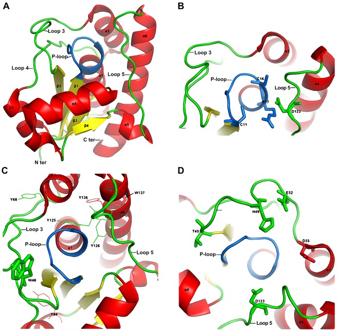
Figure 5.
(A) Cartoon structure of Tt1001 protein. α-helix, β-sheet and loops were colored in red, yellow and green, respectively. P-loop was highlighted in marine. (B) Essential residues Cys11, Cys16, Arg17 and Asp123 were showed as sticks. (C) Tryptophan and tyrosine residues were showed as sticks and lines, respectively. (D) Polar and charged residues located on loop 3 and loop 5 are showed as sticks. All the structures are prepared by PyMOL (DeLano, Warren L., The PyMOL Molecular Graphics System (2008). DeLano Scientific, California, USA).

