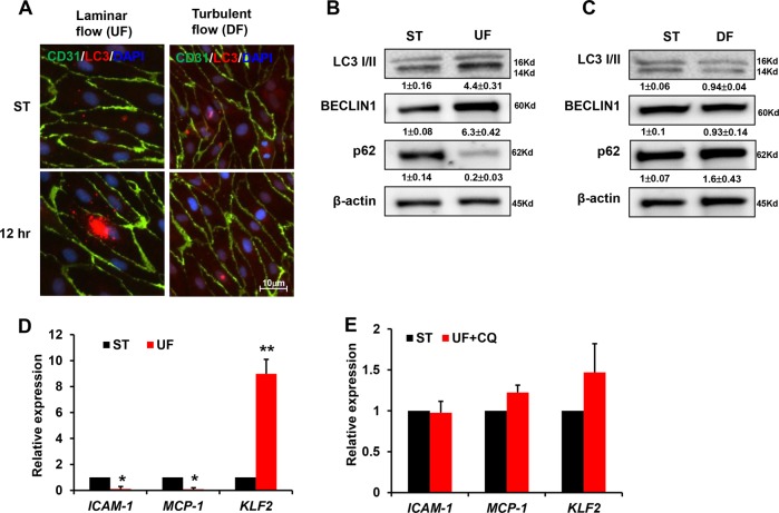
Fig. 3. Laminar flow induces autophagy.
a HUVECs were exposed to shear stress for 12 h and then subjected to immunofluorescence staining. Endothelial cells were labeled with anti-CD31 antibody. LC3 puncta indicated that UF significantly increased the number of autophagosomes in comparison with DF. Representative images from at least three independent experiments are shown. b, c Western blot analysis was performed to show the expression of BECLIN, LC3II/ LC3I, and p62 under UF or DF. Numbers under the blots were mean ± SD of three biologically independent experiments, and the first lane (ST) was served as relative control. HUVEC cells were treated with UF (d) or (e) with autophagy inhibitors, real-time PCR was performed to determine the expression of ICAM1, MCP-1, and KLF2. Data represent mean ± SD of three independent experiments (**P < 0.01, *P < 0.05, vs. the control).

