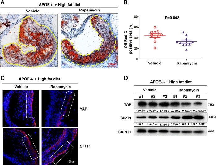
Fig. 7. Rapamycin attenuates atherosclerotic progression in mice.
a Oil Red O staining of mice aortas in the ApoE−/− mice fed a diet supplemented with cholesterol (Vehicle group) or with cholesterol plus Rapamycin (Rapamycin) for 8 weeks, and yellow dashed line indicates the size of plaque area as a percentage of total area. b Quantification of plaque size, Oil Red O-positive area in Vehicle group (n = 10) and rapamycin group (n = 10). Data are mean ± SEM. c Immunofluorescence staining for YAP and SIRT1 proteins in the atherosclerotic vessels from the vehicle group and rapamycin group. Nuclei are counterstained with DAPI (blue). White frame indicates endothelia membrane. Representative images are shown, n = 10. d Western blotting showed the total YAP protein level and SIRT1 level in tissues from vehicle group and rapamycin group. Three samples of each group were shown. Numbers under the blots were mean ± SD of three biologically independent experiments, and the first lane was served as relative control.

