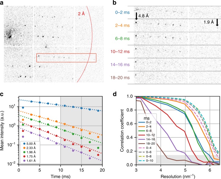
Fig. 3. Radiation damage during dose-fractionated acquisition.
a Typical diffraction pattern from a granulovirus occlusion body. The red box indicates the enlarged region in b. b Enlarged diffraction pattern section for several single frames from the dose-fractionated movie stack, each of 2 ms duration. The integration time of each frame relative to the beam first hitting the crystal is specified. Note the fading of the diffraction spots, especially at high resolutions. The first shot is affected by residual beam motion and hence has a shorter effective integration time and shows blurring artefacts. c Mean intensity of Bragg reflections for different resolution shells as a function of delay time, and exponential fit lines, where the first time point has been excluded from the fit. The shaded area corresponds to delay times beyond 10 ms, which have been excluded from our data analysis. d Resolution-dependent correlation coefficients CC1/2 shown from 3.33 to 1.55 Å resolution. Solid lines correspond to single movie frames as in b. Dashed lines correspond to images that were cumulatively summed over several frames. The shaded area corresponds to values CC1/2 <0.143, where data falls below the resolution cut-off at CC* = 0.5.

