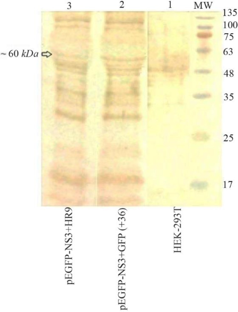
Figure 6.

Western blot analysis of the fused NS3-GFP expressed in HEK-293T cells at 48 hr after transfection using an anti-GFP polyclonal antibody (Abcam); Lane 1: un-transfected cells, Lane 2: transfected cells with +36 GFP/NS3 DNA at N/P ratio of 10:1, Lane 3: transfected cells with HR9/NS3DNA complex at N/P ratio of 5:1. MW is the molecular weight marker (10–180 kDa, Fermentase). The dominant band of ∼60 kDa was detected in transfected cells with these complexes.
