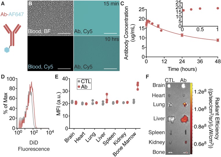
Fig. 3.
Mouse antibodies remain in circulation for an extended time. (A) Schematic of a fluorescently labeled mouse anti-human CD45 antibody. (B) Representative phase-contrast and fluorescence images of blood with or without fluorescently labeled antibodies at different concentrations in blood. (C) Circulation half-life measurements after IV administration of fluorescently labeled antibodies using quantitative microscopy (n = 3 mice per group per time point). Error bars represent SEM. Representative end-point (D) flow cytometry histograms from homogenized livers, (E) MFI values for homogenized organs (n = 3 to 4 mice per group per time point; error bars represent SEM), and (F) IVIS analyses of NP uptake in various tissues. (Scale bars, 100 μm.)

