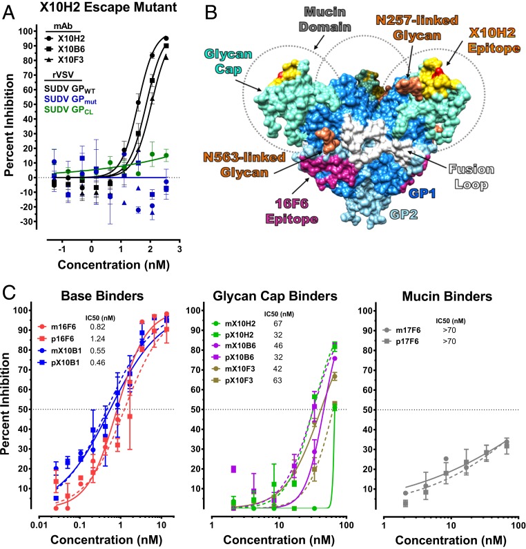
Fig. 3.
In vitro characterization of SUDV-specific mAbs. (A) rVSV-SUDV GP escape mutants to X10H2 were selected by growing the virus in the presence of IC90 concentrations of X10H2. Escape variants were then sequenced to identify GP mutations. Neutralizing activity of X10H2, X10B6, and X10F3 against wild-type rVSV-SUDV GP (GPWT) and escape mutant rVSV-SUDV GP (GPmut) was evaluated. Neutralizing activity of X10H2 against cleaved rVSV-SUDV GP (GPCL) was also evaluated. Data points represent the mean ± SEM of two replicate assays, each having two technical replicates. (B) SUDV GP structure with labeled subunits, functional elements, and known antibody epitopes. (C) Neutralizing activity of plant (p, dotted line) and mammalian (m, solid line) mAbs were evaluated by microneutralization assay. Serially diluted mAbs were mixed with SUDV prior to adding to Vero cells at an MOI of 0.5. Cells were fixed 48 h after infection and immunostained with SUDV-specific antibody and fluorescently labeled secondary antibody. Cells were imaged and the percent of infected cells was determined using an Operetta and Harmonia Software. Data are presented as percent inhibition relative to untreated, infected control cells and represent the mean ± SEM of two replicate assays, each having two technical replicates.

